At What Age Does Bone Loss Start? A Comprehensive Guide
Peak bone mass arrives around age 30, but some bone loss starts earlier than most people expect. See how loss differs by bone type and sex, what speeds it up, and when a DEXA scan makes sense.

When Bone Loss Usually Starts
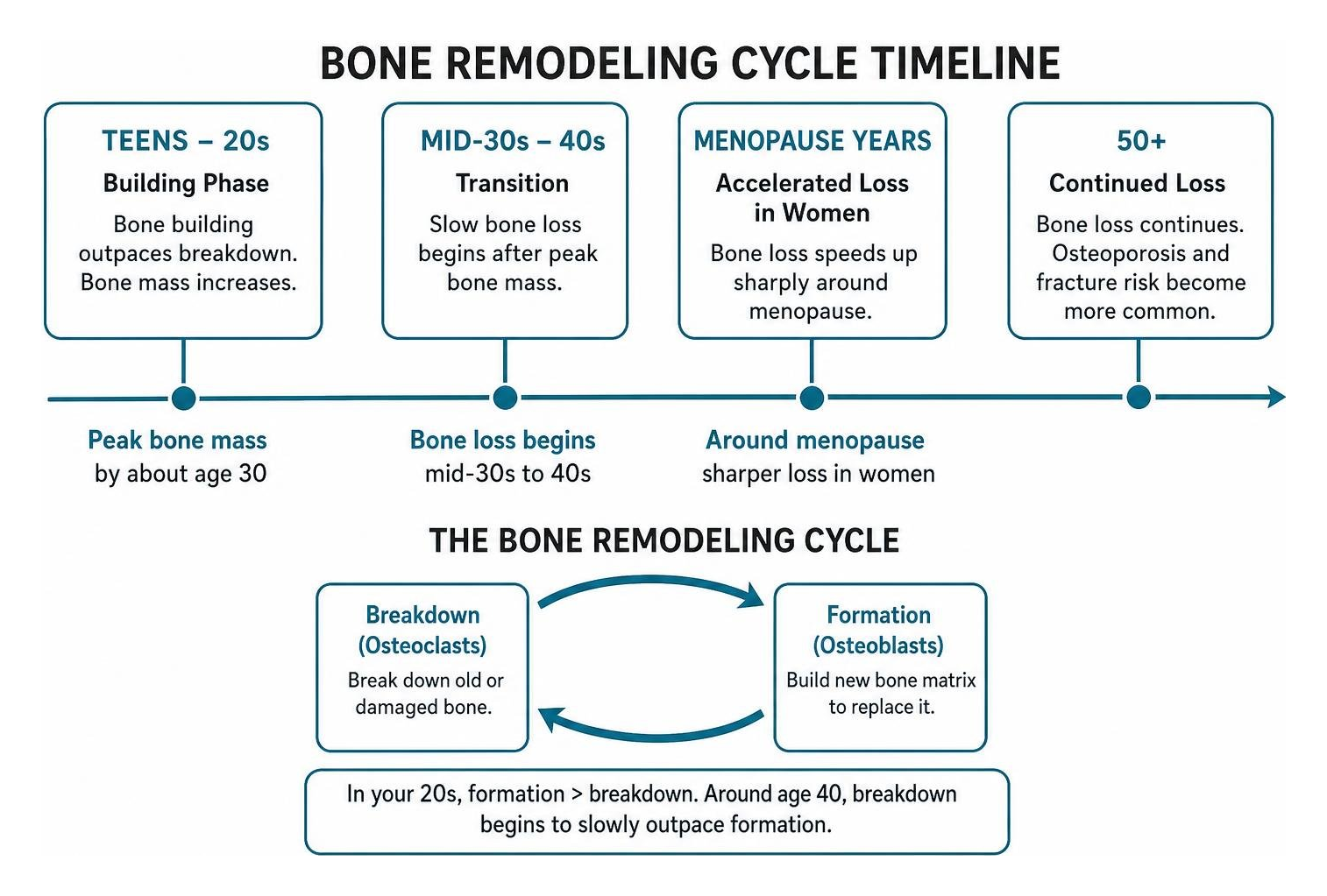
Most people reach peak bone mass by about age 30, and slow bone loss typically begins in the mid-30s to 40s, although a longitudinal Mayo Clinic study found that loss of the spongy (trabecular) inner bone starts even earlier, in young adulthood. While the spongy inner part of bone can begin thinning earlier in adulthood, loss speeds up sharply around menopause in women and progresses more gradually in men.
This does not mean osteoporosis starts at the same age. Osteoporosis and fracture risk become far more common later in life, especially after age 50. Bone is constantly being broken down and rebuilt. Over time, breakdown can outpace rebuilding.
For adults 65 and older, the key point is simple: bone loss may have started decades before a dual-energy X-ray absorptiometry (DEXA) scan, a low-dose X-ray test that measures bone density, detects low bone mass. A history of early menopause, long-term steroid use, smoking, low body weight, or family fractures can move that timeline earlier. Screening is generally recommended for women age 65 and older and men age 70 and older, with earlier testing for people with risk factors.
At What Age Does Bone Loss Start in Men and Women?
Bone loss can begin in the mid-30s, but it often becomes more noticeable in the 40s, after peak bone mass is reached around age 30.
Peak bone mass refers to the maximum amount of bone tissue a person builds during growth. Most people achieve this peak by age 30. After reaching this peak, your skeleton maintains a balance between breaking down old bone and making new bone. This continuous cycle is called bone remodeling.
Two main cell types control this process. Osteoclasts are specialized cells that break down old or damaged bone tissue. Osteoblasts are cells that build new bone matrix to replace it. In your 20s, osteoblasts build bone faster than osteoclasts break it down. Around age 40, this balance shifts. Osteoclast breakdown begins to slowly outpace osteoblast formation. This normal age-related decline is gradual during midlife.
At What Age Does Bone Loss Start in Trabecular vs Cortical Bone?
Trabecular bone loss starts earlier, often during young adulthood in the 20s and 30s, while cortical bone loss typically begins around age 40 in women and later in life for men.
The human skeleton is made of two main types of bone tissue:
- Trabecular bone: This is the spongy, honeycomb-like inner meshwork found inside vertebrae and at the ends of long bones like the hip. It has a high surface area and turns over quickly. Research shows that trabecular thinning begins quietly in young adulthood. About 37 percent of lifetime trabecular loss in women and 42 percent in men occurs before age 50.
- Cortical bone: This is the dense, solid outer shell that forms the protective shaft of long bones. Cortical bone remains stable longer. It rarely shows significant thinning until perimenopause in women and around age 75 in men.
The Menopause Transition and Accelerated Bone Loss
Menopause causes a sharp spike in bone loss because ovarian production of estrogen drops rapidly.
Estrogen is a reproductive hormone that plays a major protective role in the skeleton. It naturally limits how long osteoclasts live, keeping bone breakdown in check. When estrogen levels drop during perimenopause and menopause, osteoclasts live longer and break down bone much faster. Understanding how estrogen affects osteoblasts and osteoclasts helps explain why this shift happens so quickly.
According to the American Academy of Orthopaedic Surgeons, women can lose up to 40 percent of their spongy trabecular bone and 10 percent of their cortical bone in the decade after menopause. Learning more about how menopause affects bones gives clearer insight into why fracture risks rise after age 50.
How Bone Density Changes Decade by Decade
Bone density follows a clear progression across the lifespan, moving from peak strength in early adulthood to gradual thinning in later decades. For additional age-based guidance, the American Academy of Orthopaedic Surgeons offers healthy bones lifespan guidance.
- 20s: Bone mineral accrual finishes. Most individuals achieve peak bone mass between ages 25 and 30.
- 30s: Bone remodeling stays mostly balanced. Very early, subtle trabecular thinning starts in the spine and hips, but overall density remains stable.
- 40s: A slow, steady decline begins in cortical bone. Breakdown starts slightly outpacing formation in both sexes.
- 50s: Women enter an accelerated loss phase due to menopause, losing 1 to 2 percent of bone density annually for several years. Men continue a slow, gradual loss.
- 60s and 70s: The rapid postmenopausal loss in women slows down to match the gradual age-related loss seen in men. Secondary factors like decreased vitamin D synthesis and reduced activity can increase fracture risk for both sexes.
| Decade of Life | Primary Bone Remodeling Activity | Trabecular Bone Status | Cortical Bone Status |
|---|---|---|---|
| 20s | Formation equals or exceeds resorption | Reaches peak density | Reaches peak density |
| 30s | Balanced remodeling | Onset of subtle, slow thinning | Completely stable |
| 40s | Resorption begins to exceed formation | Ongoing gradual loss | Onset of slow thinning |
| 50s | Rapid resorption in women; steady in men | Accelerated loss in women | Noticeable thinning in women |
| 60s+ | Steady age-related loss in both sexes | Continued gradual decline | Progressive porosity |
Factors That Accelerate Bone Loss Before Age 50
Several lifestyle and medical factors can cause bone loss to start earlier or progress faster before midlife.
Reviewing osteoporosis facts and bone health tips highlights how daily choices and medical history influence skeletal strength:
- Family history: Having a parent who suffered a hip fracture increases your risk of lower peak bone density and earlier bone loss.
- Prolonged corticosteroid use: Medications like prednisone taken for more than three months suppress osteoblasts and reduce calcium absorption.
- Low body weight: Being underweight - generally a body mass index below about 19-20 is linked to smaller, thinner bones and provides less mechanical loading to stimulate bone retention, which raises the risk of osteoporosis.
- Physical inactivity: Bones adapt to the forces placed on them. Lack of resistance exercises leaves bones without a signal to maintain mineral density.
- Tobacco and excess alcohol: Smoking damages osteoblast function, while consuming more than two alcoholic drinks daily impairs calcium balance.
Key Factors Influencing at What Age Bone Loss Starts
Certain medical conditions trigger secondary bone loss decades ahead of normal aging.
Surgical removal of both ovaries causes immediate surgical menopause, triggering immediate estrogen loss in younger women. Understanding what bone changes menopause creates can help individuals prepare for these shifts. Malabsorption disorders like celiac disease or Crohn's disease block the gut from absorbing dietary calcium and vitamin D. In men, hypogonadism (abnormally low testosterone levels) leads to low levels of active estrogen, accelerating bone breakdown in midlife.
Bone Density Screening: When to Get Tested
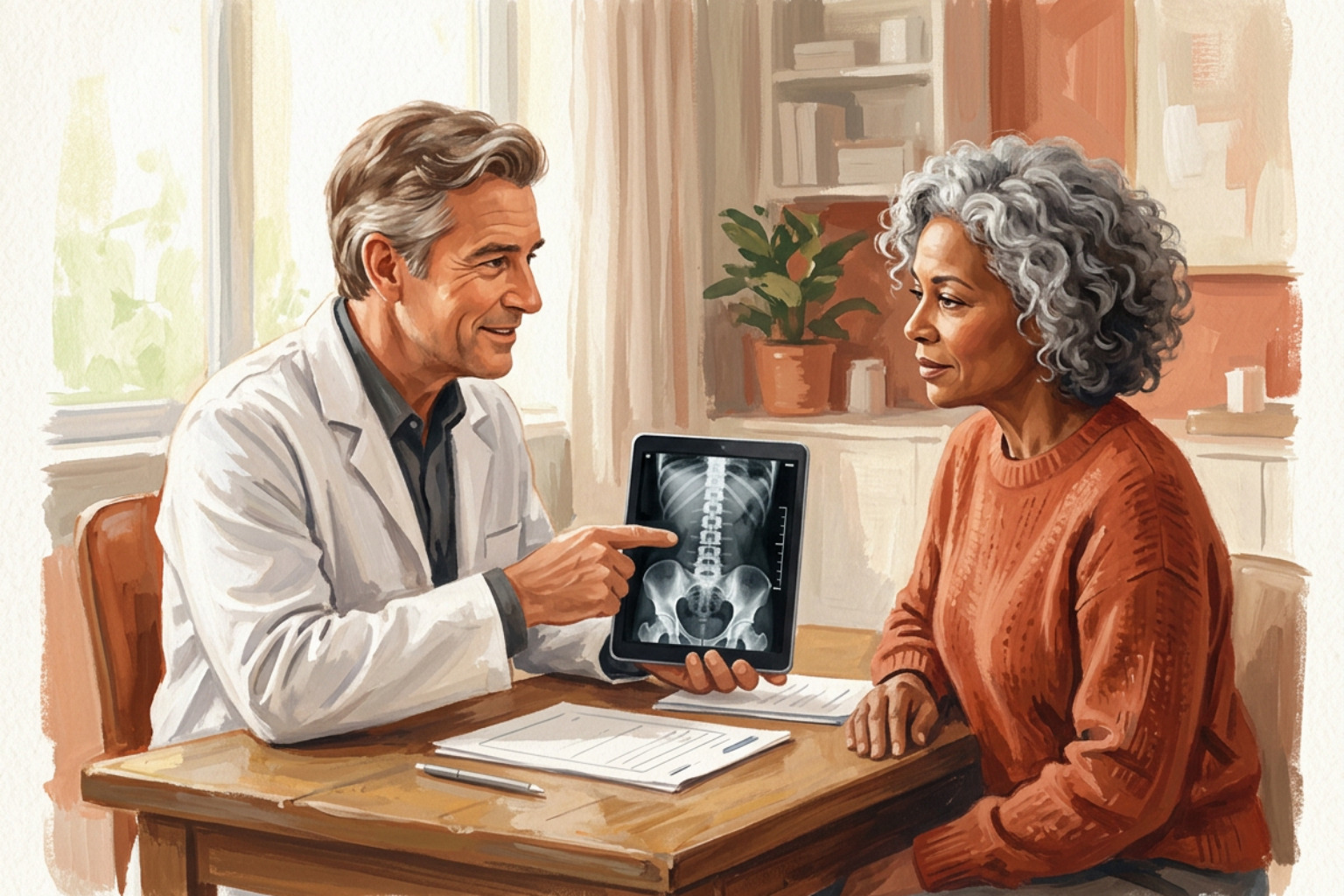
The Bone Health and Osteoporosis Foundation recommends a baseline dual-energy X-ray absorptiometry (DEXA) scan for all women aged 65 and older and all men aged 70 and older.
A DEXA scan is a low-dose X-ray test that measures bone mineral density at the hip and lumbar spine. It compares your bone density to that of a healthy young adult, producing a score called a T-score.
Earlier screening before age 50 or during perimenopause is recommended for individuals with major risk factors, such as long-term steroid therapy, a history of fragility fractures, or rheumatoid arthritis.
To interpret your scan results, review explanations of T-scores and Z-scores and check a standard T-score reference range. A T-score of -1.0 or above is normal, between -1.0 and -2.5 indicates osteopenia (low bone mass), and -2.5 or lower defines osteoporosis.
Strategies to Preserve Bone Health at Every Stage of Life
Slowing bone loss requires a consistent combination of targeted exercise, adequate nutrition, and balance training.
- Targeted resistance training: Building bone requires osteogenic loading. This means using heavy resistance training with lower repetitions and challenging weights to create mechanical tension. Prioritize compound movements that load the hip and spine, such as squats, lunges, and hip hinges. Focus on spinal extension and avoid loaded spinal flexion (bending forward under heavy weight).
- Understand exercise roles: Light activities like walking, using stretchy resistance bands, high-repetition circuits, and gentle stretching support mobility and heart health, but they do not provide enough force to stimulate bone growth.
- Progressive balance and power training: Prevent falls by practicing balance exercises using stable, braced supports (like a heavy kitchen counter or a sturdy chair placed against a wall). Progress from static balance to dynamic movement under professional guidance. Power-based movements that combine strength and speed can also help active adults react quickly to trips, but always seek professional clearance first.
- Daily calcium intake: According to the NIH Office of Dietary Supplements, women over 50 and men over 70 need 1,200 milligrams of calcium daily, ideally from foods like yogurt, fortified beverages, and dark leafy greens.
- Vitamin D3: The NIH recommends 600 to 800 International Units of vitamin D daily for older adults to aid calcium absorption in the intestine.
Frequently Asked Questions
Can you rebuild bone density after bone loss begins?
Yes, you can improve bone strength and slow loss after it begins. Targeted progressive resistance training places mechanical stress on the skeleton, signaling osteoblasts to build new bone tissue. For individuals with significant bone loss or osteoporosis, physicians may recommend medications that slow down osteoclasts or stimulate osteoblasts. Review clinical guidance on when to start osteoporosis treatment to see when prescription therapy is needed alongside exercise.
How do I know if I am losing bone mass without symptoms?
Bone loss is silent and does not cause pain on its own. Many people only discover they have low bone mass after a minor fall results in a break. Subtle signs can include a loss of height of an inch or more, or a gradual rounding of the upper back. Routine DEXA screening remains the only reliable way to detect bone loss early. Understanding your T-score helps make sense of how scans detect these silent changes.
Does bone loss occur at the same rate in men and women?
No. Bone loss does not follow the same pattern in men and women. Women often lose bone more quickly during the menopause transition and early postmenopause, when lower estrogen can speed up bone resorption. That faster phase may last several years before slowing again. Men usually have a more gradual, steady decline that often becomes noticeable around midlife. In older men, faster bone loss has been associated with lower levels of biologically active sex steroids, including estradiol, which is made in part from testosterone. Over a lifetime, men typically lose less total bone than women.
Conclusion
Bone loss is a natural part of aging that begins decades before fractures occur. While age-related thinning starts in midlife, targeted strength and balance training can help protect your mobility and skeletal health over time. Groove Health supports older adults with personalized, home-based exercise plans designed by physical therapists and overseen by physicians to help build bone strength and prevent fractures.
Works Cited
- Riggs BL, Melton LJ, Robb RA, et al. "A population-based assessment of rates of bone loss at multiple skeletal sites: evidence for substantial trabecular bone loss in young adult women and men." Journal of Bone and Mineral Research, 2008.
- Riggs BL, Melton LJ 3rd, Robb RA, et al. "Population-based study of age and sex differences in bone volumetric density, size, geometry, and structure at different skeletal sites." Journal of Bone and Mineral Research, 2004.
- American Academy of Orthopaedic Surgeons. "Healthy Bones at Every Age." OrthoInfo, accessed October 1, 2026.
- Bone Health & Osteoporosis Foundation. "Bone Density Test, Osteoporosis Screening & T-score Interpretation." Bone Health & Osteoporosis Foundation, accessed October 1, 2026.
- National Institutes of Health Office of Dietary Supplements. "Calcium - Health Professional Fact Sheet." NIH Office of Dietary Supplements, accessed October 1, 2026.
- National Institutes of Health Office of Dietary Supplements. "Vitamin D - Health Professional Fact Sheet." NIH Office of Dietary Supplements, accessed October 1, 2026.
This content is for informational purposes only and does not constitute medical advice. Consult a qualified healthcare professional for diagnosis and treatment. If you are on Medicare and interested in a personalized bone health program, you can check your eligibility at groovehealth.com.