
Postmenopausal Bone Loss: A Scientific Guide to Estrogen and Bone Density
Bone breaks down faster than it rebuilds once estrogen falls. This guide explains the biology, how DEXA and FRAX measure risk, and the treatment, nutrition, and exercise steps that help protect bone strength.

Why Estrogen Loss Changes Bone Strength After Menopause
Postmenopausal bone loss science explains why bones can become weaker more quickly after menopause. As estrogen levels fall, the body speeds up bone breakdown more than bone rebuilding. This can lower bone mineral density, thin the inner structure of bone, and raise the chance of breaks from a fall or everyday force.
Bone is living tissue. It is always being removed and rebuilt. After menopause, falling estrogen shifts this balance in two ways: osteoclasts resorb bone faster, and osteoblasts often cannot replace it quickly enough. Estrogen loss can also shorten osteoblast survival. In the Study of Women's Health Across the Nation (SWAN), a U.S. cohort, lumbar spine bone density fell about 10.6% over ten years, and about 7.4% of that loss happened from one year before to two years after the final menstrual period.
That does not mean a fracture is inevitable. A DEXA scan, an X-ray that measures bone density, can help show whether bone loss needs closer attention. The National Institute of Arthritis and Musculoskeletal and Skin Diseases explains osteoporosis and its diagnosis in more detail.
This guide covers the biology behind bone loss, what density tests can and cannot tell you, and the practical medical, nutrition, activity, and fall-prevention steps that may help protect bone strength. For a closer look at how estrogen affects the cells that build and break down bone, see how estrogen affects osteoblasts and osteoclasts.
How Menopause Changes Bone Cells: Cellular and Molecular Drivers
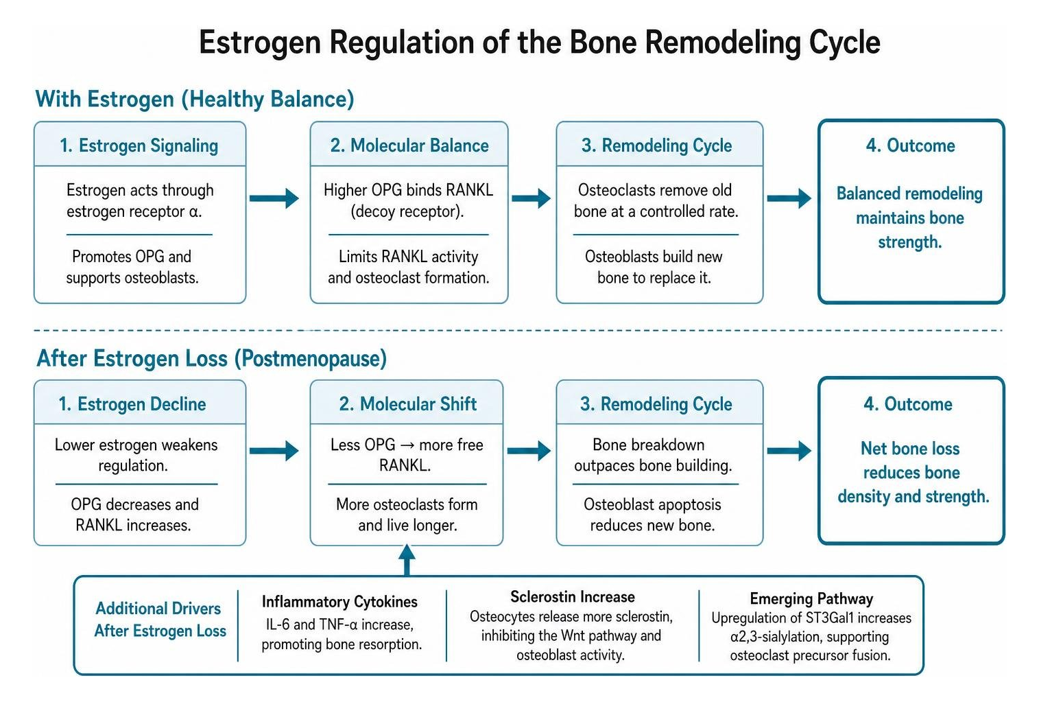
Understanding the cellular basis of how menopause affects bones begins with the balance between two primary cell types: osteoclasts (cells that break down old bone) and osteoblasts (cells that build new bone). In healthy adult bone, these cells work in closely linked pairs to replace old bone tissue with new mineralized bone. Estrogen serves as a natural brake on bone breakdown while supporting the lifespan of bone-building cells. When ovarian estrogen production drops, this regulatory control weakens, and the remodeling cycle falls out of balance.
How Estrogen Regulates Bone Remodeling
At the molecular level, estrogen controls the chemical signaling between bone-building and bone-clearing cells. Under normal conditions, estrogen promotes protective signaling proteins that prevent bone-clearing cells from becoming overactive. When estrogen levels fall, the signals that trigger bone breakdown increase. This shift speeds up the creation of bone-resorbing cells and extends their active lifespan.
At the same time, the loss of estrogen signaling leads to early cell death among bone-building cells. The Endocrine Society on osteoporosis notes that women begin to lose bone at an even faster rate after menopause. This mismatch is generally described as deeper resorption pits, with less replacement bone formed in each remodeling cycle.
Cellular Changes and Inflammatory Signaling
Estrogen decline also allows an increase in circulating inflammatory signals throughout the body. These natural chemical messengers further encourage bone breakdown cells to mature and dissolve mineralized tissue. Concurrently, resting bone cells release higher levels of inhibitory signals that block bone-forming pathways. Together, these combined cellular shifts tip the entire skeletal environment toward persistent, measurable bone loss.
Structural Bone Changes: Bone Density, Periosteal Apposition, and Strength Index

The cellular imbalance caused by menopause alters both the inner meshwork and outer shell of the skeleton. Trabecular bone, the spongy lattice found inside the vertebrae and hip, undergoes thinning and loss of structural connectivity. Cortical bone, the dense outer layer of long bones, experiences increased porosity and endocortical resorption, meaning bone is removed from the inner surface facing the marrow cavity.
Periosteal Apposition vs. Medullary Expansion
As estrogen levels decline, the forearm bones studied undergo a natural geometric shift. The inner marrow space widens through medullary expansion. To help counteract this loss of inner material, the outer surface of the bone slowly adds new tissue through a process called periosteal apposition. In a 15-year follow-up of 108 women, the medullary diameter of the distal radius (the wrist end of the forearm) widened by about 1.1% per year after menopause, while the periosteal diameter increased by about 0.7% per year.
This outer expansion increases the section modulus, a geometric measurement of resistance to bending. While this structural adaptation helps maintain some mechanical strength, research on what bone changes menopause triggers indicates that periosteal growth only partly preserves bone strength as inner bone density falls.
Beyond BMD: The Bone Strength Index
Bone mineral density (BMD) measures the amount of mineral in a two-dimensional area, but it does not capture the three-dimensional geometry of the skeleton. A composite strength index combines bone mineral apparent density with cross-sectional dimensions to evaluate how bone handles mechanical load.
In that same study, a strength index combining bone density with bone size predicted wrist fractures: each 1-standard-deviation lower baseline value was associated with a risk ratio of 3.8 (95% CI 1.8 to 8.0) for distal radius fracture. That suggests structural architecture may matter alongside DEXA T-score fracture risk assessments, although this was a small study at a single skeletal site and needs confirmation.
Risk Factors, Demographics, and Diagnostic Evaluation
| Diagnostic and Assessment Tool | Primary Measurement | Clinical Role in Evaluation |
|---|---|---|
| DEXA Scan (DXA) | Areal bone mineral density (g/cm2) | Gold standard for diagnosing osteopenia and osteoporosis via T-scores |
| T-Score | Standard deviations from young adult mean | Classifies bone mass: normal is -1.0 or higher, osteopenia is between -1.0 and -2.5, osteoporosis is -2.5 or lower |
| FRAX Calculator | 10-year probability of major osteoporotic fracture | Integrates BMD with clinical risk factors to guide treatment thresholds |
| Trabecular Bone Score (TBS) | Pixel variation from lumbar spine DEXA | Evaluates microarchitectural texture to refine fracture risk estimation |
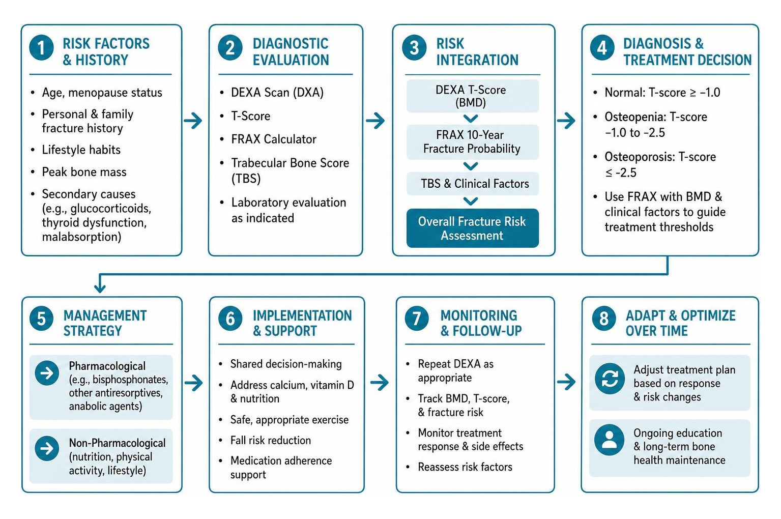
A complete clinical evaluation for osteoporosis includes assessing lifestyle habits, personal and family fracture history, and evaluating peak bone mass attained earlier in life. Clinicians also check for secondary causes of bone thinning, such as chronic glucocorticoid use, thyroid dysfunction, and malabsorption disorders.
Racial, Ethnic, and Nutritional Disparities in Bone Loss
Bone density ranges and fracture probabilities vary across different populations. Public health data from the Bone Health and Osteoporosis Foundation estimate that about 54 million Americans have osteoporosis or low bone mass, and that about one in two women and up to one in four men aged 50 and older will break a bone due to osteoporosis.
Nutritional factors also differ across demographic groups. High rates of lactose intolerance can limit dietary calcium consumption from dairy products, and vitamin D insufficiency remains widespread globally. However, healthcare organizations emphasize that individual fracture risk varies widely and should always be assessed on personal clinical factors rather than racial or ethnic categories alone.
Clinical Diagnosis: DEXA, T-Scores, and the FRAX Tool
Dual-energy X-ray absorptiometry (DEXA) remains the primary diagnostic method for assessing bone health. When interpreting DEXA scan results, clinicians evaluate the T-score, which compares a patient's bone density to that of a healthy young adult reference population.
According to standard diagnostic criteria, a T-score of -1.0 or higher is considered normal. A T-score between -1.0 and -2.5 falls into the low bone mass range, as shown on a standard T-score osteopenia range chart. A score of -2.5 or lower defines osteoporosis. To translate these numbers into real-world terms, doctors use the FRAX tool, which calculates the 10-year probability of a major osteoporotic break by combining bone density with factors like age, body weight, smoking, and prior fractures.
Evidence-Based Management: Pharmacological and Non-Pharmacological Interventions
Managing postmenopausal bone loss involves a structured approach combining medical management, adequate nutrition, and safe physical activity. Guidelines help clinicians determine when to start osteoporosis treatment based on personal fracture risk and T-scores. All prescription medications and hormone therapies are clinician-directed treatments prescribed by personal physicians. Groove Health is a Medicare covered bone health program for adults 65 and older that pairs each member with a physician and a physical therapist who designs a personalized exercise plan.
Medical Treatments for Postmenopausal Bone Loss
Medical treatments for bone loss fall into two main categories: antiresorptive medications (which slow breakdown) and bone anabolic agents (which stimulate new growth).
- Bisphosphonates: Drugs such as alendronate (Fosamax), risedronate (Actonel), and annual zoledronic acid infusions (Reclast) bind to bone minerals and reduce the activity of bone-resorbing cells. In the HORIZON trial, three annual infusions of zoledronic acid (Reclast) reduced the risk of morphometric vertebral fracture by 70% over three years compared with placebo (3.3% vs. 10.9%).
- Targeted Breakdown Inhibitors: Denosumab (Prolia) is a targeted injection administered every six months to slow the development of bone-clearing cells. Because stopping denosumab(Prolia) can trigger a rapid rebound surge in bone turnover, clinicians often transition patients to a bisphosphonate if treatment is paused.
- Anabolic Agents: Medications such as teriparatide (Forteo) and abaloparatide (Tymlos) stimulate bone-building cells to produce new bone tissue, typically reserved for individuals at very high fracture risk.
- Hormone Therapy: Estrogen or combined hormone therapy, such as estradiol-based therapy or conjugated estrogens with progestin (Prempro), helps preserve bone density during the menopause transition, though potential cardiovascular and other risks require individualized medical evaluation.
Nutrition, Lifestyle, and Safety Considerations
Adequate nutrition provides the raw materials necessary for bone remodeling. Ensuring adequate intake of essential nutrients for bone health, particularly calcium and vitamin D, supports ongoing bone mineralization. According to the National Institutes of Health (NIH) Office of Dietary Supplements, the calcium RDA is 1,200 mg per day for women older than 50, and the vitamin D RDA is 600 IU through age 70 and 800 IU from age 71, which supports intestinal calcium absorption.
Adequate dietary protein, often around 0.8 to 1.2 grams per kilogram of body weight daily, helps maintain the bone collagen matrix and support muscle strength. Beyond nutrition, structured physical activity can support bone by exposing the skeleton to safe mechanical loading, especially through weight-bearing movement, resistance-based activity, and balance-focused training. The science is explained further in how exercise builds bones.
Long-term medication safety requires clinical oversight. Rare side effects such as osteonecrosis of the jaw and atypical femoral fractures are managed with routine dental care and clinician monitoring. An ASBMR task force found that the risk of atypical femoral fracture rises with treatment duration, and suggests reassessing risk after 5 years of oral or 3 years of intravenous bisphosphonate treatment, with longer treatment (up to 10 years oral or 6 years intravenous) considered for women at high risk.
Frequently Asked Questions about Postmenopausal Bone Loss
How quickly does bone loss occur after menopause begins?
Bone loss is fastest from about one year before to two years after the final menstrual period. In the SWAN cohort, lumbar spine bone density fell about 7.4% during those three years and then kept falling more slowly, reaching about 10.6% over ten years.
Can bone size changes completely compensate for lost bone density?
No. While periosteal apposition expands the outer diameter of bone and slightly improves resistance to bending forces, it does not fully compensate for the loss of internal trabecular connections and thinning cortical bone. Overall bone strength still declines without appropriate lifestyle and clinical interventions.
When should a postmenopausal woman begin screening and treatment?
Routine DEXA screening is widely recommended for all women aged 65 and older, and earlier for postmenopausal women with risk factors such as prior fractures, low body weight, or medications that cause bone thinning. According to Medicare.gov coverage rules for bone mass measurements, Medicare Part B covers bone density measurements once every 24 months (or more often if medically necessary) with zero out-of-pocket cost when the provider accepts assignment, though specific coverage rules may vary by plan type. Treatment decisions are made in consultation with a physician when T-scores reach the osteoporotic range or when FRAX scores indicate high fracture probability.
Key Takeaways: Protecting Bone Strength After Menopause
The scientific link between menopause and bone loss centers on the essential role of estrogen in regulating skeletal remodeling. When estrogen decline removes the natural check on osteoclasts, bone resorption outpaces bone formation, leading to decreased bone density, microarchitectural thinning, and higher fracture susceptibility.
While natural geometric adjustments like periosteal expansion provide limited structural support, preserving long-term skeletal strength requires a comprehensive approach. Routine DEXA screening, FRAX risk estimates, targeted nutrition, progressive strength training, appropriate impact training, and clinician-guided pharmacotherapy can work together to protect bone health throughout aging.
Early evaluation and evidence-based care help maintain independent mobility and significantly reduce the chance of debilitating fractures over time.
Works Cited
- Greendale GA, Sowers M, Han W, et al. "Bone mineral density loss in relation to the final menstrual period in a multiethnic cohort: results from the Study of Women's Health Across the Nation (SWAN)." Journal of Bone and Mineral Research, 2012.
- Ahlborg HG, Johnell O, Turner CH, Rannevik G, Karlsson MK. "Bone loss and bone size after menopause." New England Journal of Medicine, 2003.
- Black DM, Delmas PD, Eastell R, et al. "Once-yearly zoledronic acid for treatment of postmenopausal osteoporosis." New England Journal of Medicine, 2007.
- Adler RA, El-Hajj Fuleihan G, Bauer DC, et al. "Managing Osteoporosis in Patients on Long-Term Bisphosphonate Treatment: Report of a Task Force of the American Society for Bone and Mineral Research." Journal of Bone and Mineral Research, 2016.
- Bone Health & Osteoporosis Foundation. "What is Osteoporosis and What Causes It?" Bone Health & Osteoporosis Foundation, 2024.
- National Institute of Arthritis and Musculoskeletal and Skin Diseases. "Osteoporosis: Diagnosis, Treatment, and Steps to Take." NIAMS, National Institutes of Health, accessed October 7, 2026.
- Endocrine Society. "Osteoporosis." Endocrine Society Hormone Health Library, accessed October 7, 2026.
- National Institutes of Health Office of Dietary Supplements. "Calcium - Health Professional Fact Sheet." NIH Office of Dietary Supplements, accessed October 7, 2026.
- National Institutes of Health Office of Dietary Supplements. "Vitamin D - Health Professional Fact Sheet." NIH Office of Dietary Supplements, accessed October 7, 2026.
- Centers for Medicare & Medicaid Services. "Bone mass measurements." Medicare.gov, accessed October 7, 2026.
This content is for informational purposes only and does not constitute medical advice. Consult a qualified healthcare professional for diagnosis and treatment. If you are on Medicare and interested in a personalized bone health program, you can check your eligibility at groovehealth.com.