
The Ins and Outs of Trabecular Bone Loss
Trabecular bone loss weakens the spongy bone inside your spine, hip, and wrist, and a standard DXA scan can miss it. See what drives it, which tests go deeper, and how to protect bone strength.

Why Understanding Trabecular Bone Loss Matters
Understanding trabecular bone loss means knowing that the spongy inner network of bone can become thinner, less connected, and weaker over time. This may raise the chance of a broken bone, especially in the spine, hip, or wrist, even when a standard bone density test does not show the full picture.
Trabecular bone, also called cancellous bone, works like a tiny internal scaffold. Osteoporosis can thin its supporting plates, widen its spaces, and break links between them. Those structural changes can reduce bone strength and make falls more dangerous.
Bone density still matters, but it is only one part of fracture risk. This guide explains what happens inside trabecular bone, why loss occurs, and how clinical testing, treatment, movement, and fall prevention may help protect skeletal strength. Readers who want a broader overview can start with Understanding Osteoporosis.
What Is Trabecular Bone and Why Is It Vital for Skeletal Strength?
Trabecular bone is the light, porous bone tissue found inside the ends of long bones and within the vertebrae of the spine. It provides internal support and absorbs physical shocks during daily movement.
The adult skeleton contains two main types of bone tissue: cortical bone and trabecular bone. Cortical bone forms the dense outer shell, making up about 80% of total skeletal mass. Trabecular bone accounts for the remaining 20%, but it provides roughly 80% of the internal bone surface area. Because of this vast surface area, trabecular tissue is metabolically active and undergoes continuous remodeling to repair everyday microdamage and balance calcium levels in the body.
| Feature | Cortical Bone | Trabecular Bone |
|---|---|---|
| Location | Outer protective shell of bones, shafts of long bones | Interior of vertebrae, pelvis, and ends of long bones |
| Structure | Dense, solid, organized in osteons | Porous, 3D honeycomb network of plates and rods |
| Surface Area | Lower surface-to-volume ratio | High surface-to-volume ratio |
| Metabolic Activity | Slower turnover rate | Rapid cellular turnover and remodeling |
| Primary Function | Torsional and bending resistance | Shock absorption and load distribution |
Trabecular Architecture and Plate-to-Rod Transition
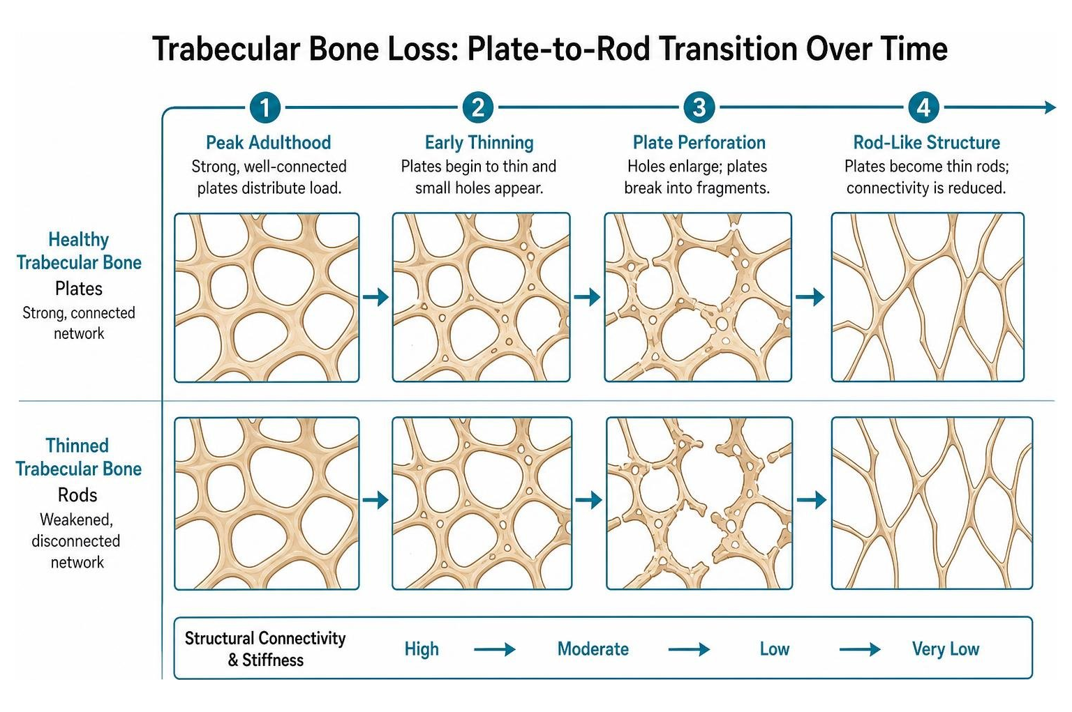
Healthy trabecular bone looks like a honeycomb made of flat structural plates interconnected by thin rods. These plates distribute mechanical forces across the joint or vertebral body.
When bone loss occurs, these flat plates thin out and develop tiny perforations. As these holes expand, the plates transform into thin, rod-like structures. This plate-to-rod transition reduces the structural connectivity of the network. Without connected horizontal struts to support vertical loads, bone stiffness drops significantly, making the bone far more vulnerable to breaking under normal daily loads.
How Trabecular Bone Changes From Your 20s Onward
Trabecular bone mass reaches its peak in early adulthood. According to the International Osteoporosis Foundation, bone mass naturally begins to decline as the natural remodeling process slowly shifts toward greater bone removal than bone replacement.
Research suggests that trabecular bone loss begins earlier than cortical bone loss, often starting quietly in a person's 20s in both men and women. In one large study, participants lost more than a third of their lifetime trabecular bone before age 50. In women, this loss often speeds up around menopause as estrogen levels fall, especially in the lumbar spine. In men, trabecular struts tend to thin steadily over time; declining sex hormones may contribute later in life, though researchers have not fully explained why the loss starts so early.
How Bone Cells Drive Trabecular Loss
Trabecular bone loss occurs when the balance between bone-resorbing cells (osteoclasts) and bone-building cells (osteoblasts) is disrupted.
Under healthy conditions, osteocytes act as internal strain sensors within the bone matrix. When everyday activities bend or compress bone tissue, osteocytes detect these mechanical signals through mechanotransduction and coordinate remodeling. Osteoclasts remove old or damaged bone tissue, and osteoblasts fill the cavity with new mineralized matrix. When osteoclast activity outpaces osteoblast formation, bone structure degrades.
Deep Resorption Pits and Trabecular Perforation
During rapid bone turnover, especially around menopause, osteoclasts can dig deeper pits into individual trabeculae. Osteoblasts normally refill these pits, but with age they often replace slightly less bone than was removed, causing gradual thinning. If a pit cuts through a thin trabecula, that connection may be permanently lost because there is no surface left to rebuild on.

When resorption cuts entirely through a thin trabecular strut, it causes an irreversible perforation. Once a trabecula is completely broken, the cellular framework is lost, meaning osteoblasts have no surface left to lay down new bone tissue. This leads to permanent loss of structural connectivity and creates localized stress concentrations around neighboring struts.
Mechano-biological Regulation and Tissue Stiffening
As trabecular struts thin, remaining bone tissue experiences altered stress distributions during daily tasks. Computational models and micro-finite element analyses show that regional structural deterioration directly impacts mechanical strength.
In trabecular-rich areas like the vertebrae, even small amounts of bone loss can lead to much larger drops in strength. That is because bone strength often falls faster than density, and losing connections between trabecular struts weakens the network more than simple thinning. At the hip, both the trabecular core and dense outer shell matter for fracture risk.
Clinical Assessment, Fracture Risk, and Diagnostic Tools
Understanding trabecular bone matters because a standard two-dimensional DXA scan measures how much mineral is in your bones, but it cannot show the three-dimensional trabecular network inside them.
Many people who experience a fragility fracture do not have T-scores in the osteoporosis range. In one study of nearly 150,000 postmenopausal women, more than 80% of those who broke a bone had T-scores above -2.5. One reason: far more people have osteopenia than osteoporosis, so that larger group accounts for many fractures. Standard scans also miss factors like falls, age, prior fractures, and internal trabecular damage.

Advanced Tests That Look Beyond Bone Density
Clinicians utilize several tools to evaluate bone quality beyond standard bone mineral density:
- Trabecular Bone Score (TBS): Software that analyzes the texture of a standard spine DXA image to estimate trabecular network quality. A higher score suggests better-preserved structure; a lower score suggests deterioration. TBS is mainly used alongside FRAX to refine fracture risk and is available at some, but not all, imaging centers.
- Vertebral Fracture Assessment (VFA): A side-view spine image taken on the same DXA machine during the same visit. It can detect symptom-free spine fractures, which may change diagnosis and treatment decisions.
- High-Resolution Peripheral Quantitative Computed Tomography (HR-pQCT): A specialized 3D scan that can show individual trabecular plates and rods, plus outer-bone porosity, at the wrist and shin. It is used mostly in research and is not widely available for routine care.
- Opportunistic Computed Tomography (CT) Screening: Using CT scans already taken for other reasons, such as chest or abdominal imaging, to estimate bone density without extra radiation. It is an emerging approach; concerning results usually lead to standard DXA testing.
While HR-pQCT provides detailed structural insights in clinical research, its use in everyday clinical practice is limited due to equipment availability and cost.
Medicare Coverage for Bone Mass Measurement
For older adults, routine screening helps detect changes in bone strength before a fracture occurs. According to Medicare.gov, Medicare Part B covers bone mass measurements (such as standard DEXA scans) once every 24 months for eligible individuals, or more frequently when medically necessary. There is generally no out-of-pocket cost if the healthcare provider accepts Medicare assignment, though exact coverage and rules may vary by plan type.
Evidence-Based Approaches to Preserve Trabecular Architecture
Protecting trabecular architecture requires a comprehensive strategy involving medical management, lifestyle modifications, and fall prevention. Groove Health is a Medicare covered bone health program for adults 65 and older that pairs each member with a physician and a physical therapist who designs a personalized exercise plan.
Evidence-based strategies to support trabecular preservation include:
- Antiresorptive Medications: Drugs like bisphosphonates and denosumab slow bone-resorbing cells, helping preserve structure and reduce fracture risk, especially in the spine and hip. Denosumab should not be stopped without a clinician-guided plan.
- Bone-Building Medications: Teriparatide, abaloparatide, and romosozumab stimulate new bone formation and may strengthen the trabecular network. They are usually time-limited and often followed by another medication to maintain gains.
- Clinician-Guided Nutrition: Enough calcium, vitamin D, and protein gives bone and muscle needed support, especially during treatment. Supplements alone have not been shown to prevent fractures for most people.
- Targeted Exercise: Supervised resistance and weight-bearing exercise can help maintain bone density and support skeletal strength.
- Comprehensive Fall Prevention: Balance and strength training can reduce falls in older adults. Because most hip fractures happen after falls, prevention is one of the most practical ways to protect fragile bones.
Frequently Asked Questions About Trabecular Bone Loss
What is the main difference between cortical and trabecular bone loss?
Trabecular bone has a much higher surface area and metabolic turnover rate than cortical bone, making it show signs of bone loss earlier during hormonal shifts or aging. While cortical bone loss typically appears as thinning and increased porosity in the outer bone wall, trabecular bone loss involves the thinning and loss of internal plates and rods.
Can perforated trabecular plates ever be restored?
Partially. Once a trabecular strut is fully severed, normal repair cannot easily bridge the gap because new bone needs an existing surface to build on. Bone-building medications, such as teriparatide, may thicken remaining struts and improve some network connections, helping strengthen bone and reduce fracture risk. These are usually reserved for people at high fracture risk. Antiresorptives, such as bisphosphonates, work differently: they help prevent new perforations rather than rebuild lost ones.
Why does standard DEXA sometimes miss early trabecular deterioration?
A standard DEXA scan provides a two-dimensional areal measurement of total mineral mass within a targeted region. It cannot differentiate between the outer cortical shell and the internal trabecular microarchitecture, nor can it show whether the internal honeycomb structure is intact or disconnected.
Key Takeaways: Protecting Your Bone Structure
Trabecular bone serves as an essential internal support structure that cushions and protects the skeleton from daily mechanical stress. When bone remodeling falls out of balance, the thinning of trabecular plates, the transition to fragile rods, and permanent strut perforations weaken the entire skeleton, raising the risk of spinal, hip, and wrist fractures.
While standard bone density testing remains a valuable foundation for skeletal assessment, understanding bone microarchitecture provides a clearer picture of bone quality. Advanced tools like Trabecular Bone Score and clinical risk evaluations help clinicians identify hidden bone fragility early.
Effective management of osteoporosis involves regular bone density monitoring, physician-prescribed medications when appropriate, balanced nutrition, and structured, safe physical activity to preserve remaining bone structure and prevent falls.
Works Cited
- Siris ES, Chen YT, Abbott TA, et al. “Bone Mineral Density Thresholds for Pharmacological Intervention to Prevent Fractures.” Archives of Internal Medicine, 2004;164(10):1108-1112.
- Silva BC, Leslie WD, Resch H, et al. “Trabecular Bone Score: A Noninvasive Analytical Method Based Upon the DXA Image.” Journal of Bone and Mineral Research, 2014;29(3):518-530.
- International Osteoporosis Foundation. “About Osteoporosis.” International Osteoporosis Foundation, accessed October 2026.
- Centers for Medicare & Medicaid Services. “Bone Mass Measurements.” Medicare.gov, accessed October 2026.
This content is for informational purposes only and does not constitute medical advice. Consult a qualified healthcare professional for diagnosis and treatment. If you are on Medicare and interested in a personalized bone health program, you can check your eligibility at groovehealth.com.