What Patients Should Know About DEXA Scan Radiation Safety and Preparation
A DEXA scan measures bone density using a very small amount of radiation. Here is what that dose actually means, why technologists skip the lead apron, and the preparation steps that keep your results accurate.
Understanding DEXA Scan Radiation Safety and Dose Levels
A dual-energy X-ray absorptiometry (DEXA) scan is a quick medical test that uses low-dose X-rays to measure bone mineral density, or bone strength. The test uses an extremely small dose of radiation, usually between 1 and 15 microsieverts (µSv) for a standard hip and spine evaluation. By comparison, everyday exposure from our natural environment delivers about 10 µSv each day, meaning a scan equals only about one or two days of ordinary living.
Ionizing radiation is a form of energy that can travel through body tissues to create detailed medical images of your skeleton. Scientists and physicians measure human radiation absorption using units called microsieverts. Radiation safety guidance from the International Atomic Energy Agency confirms that modern DEXA equipment produces minimal exposure while delivering vital diagnostic information about your bones.
| Procedure / Source | Typical Radiation Dose (µSv) | Equivalent Daily Natural Background |
|---|---|---|
| DEXA Scan (Hip & Spine) | 1 - 15 µSv | Less than 1 to 2 days |
| Full-Body DEXA Scan | 1 - 5 µSv | Less than 1 day |
| Natural Daily Background | ~10 µSv | 1 day baseline |
| Standard Chest X-Ray | 20 - 50 µSv | 2 to 5 days |
| Dual-Energy Quantitative Computed Tomography (QCT) Scan | 50 - 100 µSv | 5 to 10 days |
According to national health data from the CDC, nearly 1 in 5 women and 1 in 20 men over age 50 live with osteoporosis, a condition characterized by weak, porous bones. Identifying bone thinning early through safe screening allows you and your healthcare team to take proactive steps before a painful fracture or break occurs.
How DEXA Compares With Other Imaging Tests
DEXA scans deliver far less radiation than most standard diagnostic tests, including routine chest X-rays, mammograms, or abdominal CT scans. In clinical practice, the theoretical risk associated with this minute level of exposure is negligible compared to the significant medical benefit of diagnosing severe bone loss and preventing debilitating hip or spinal fractures.
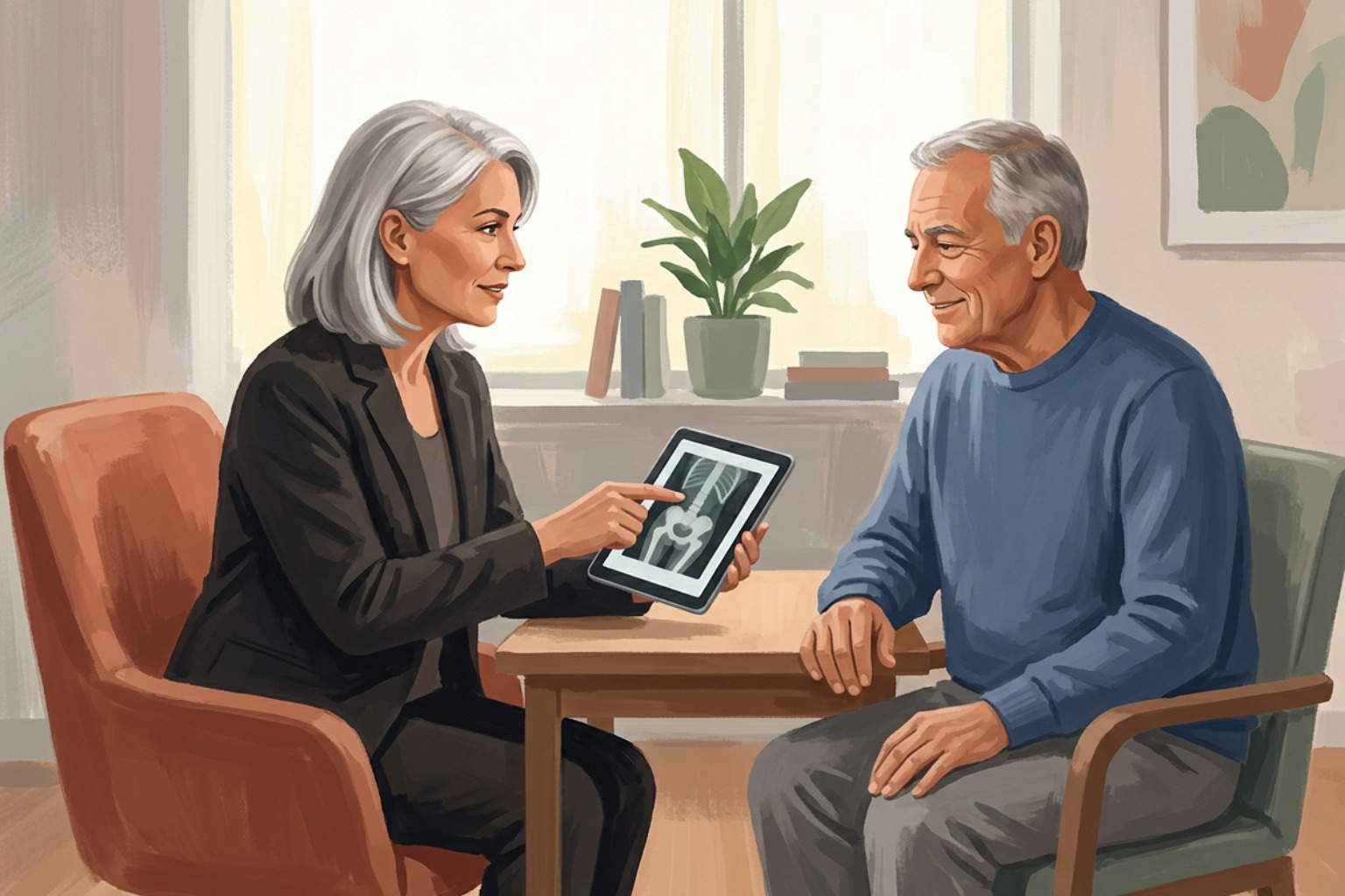
Public health guidance from the CDC points out that a bone density scan adds only a negligible amount to lifetime health risks. When your doctor reviews your scan, they look at your results to evaluate your long-term fracture risk and recommend appropriate lifestyle or medical steps. If you want to know what those numbers mean on your report, our guide on interpreting DEXA scan results walks through the details.
Scanner Technology: Pencil Beam vs Fan Beam
The specific type of scanner used by your clinic influences both the test duration and the exact radiation dose you receive during the appointment.
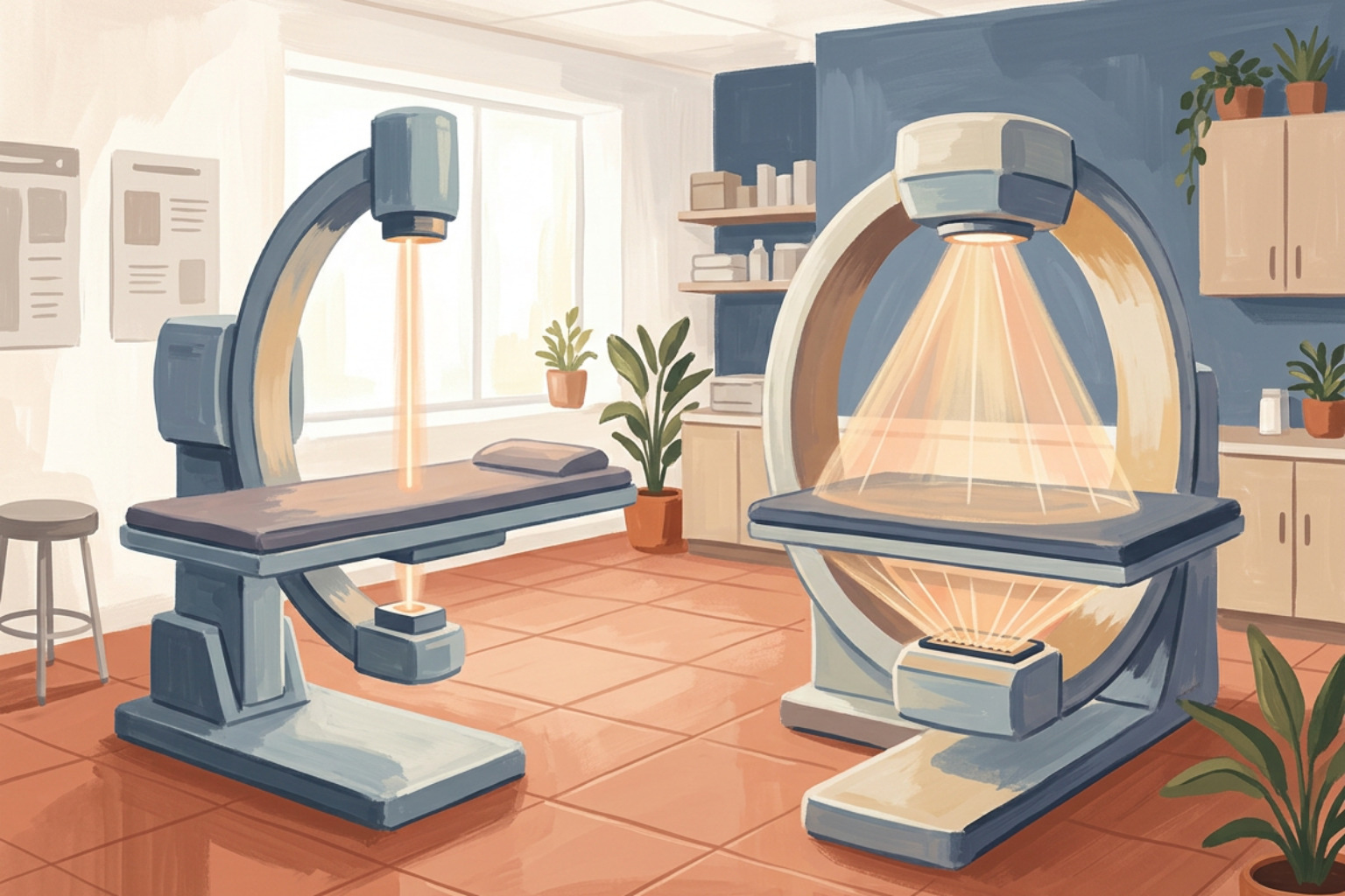
- Pencil-Beam Scanners: Older systems use a single, narrow beam of X-rays that moves back and forth in a grid pattern. These machines deliver less than 1 µSv per scan, but they take several minutes to finish each area.
- Fan-Beam and Cone-Beam Scanners: Modern imaging centers frequently use wide, fan-shaped beams that capture entire regions in one sweep. These scanners complete an image in under one minute. While the dose is slightly higher (typically between 1 and 10 µSv), it remains well below the radiation received during a routine chest X-ray and exposes surrounding tissues to minimal scatter.
Step-by-Step Preparation: How to Prepare for an Accurate Scan
Preparing for a DEXA scan requires avoiding calcium pills for 24 hours and wearing loose clothing without metal. Proper preparation ensures that the scanner reads your natural bone density rather than external objects.
When preparation steps are missed, dense items can artificially inflate your bone density numbers. This could distort your diagnostic score and make bones appear denser than they actually are. You can read our review of t-score meaning explained simply to see how these baseline scores guide medical decisions.
Medication and Dietary Restrictions Before Your Appointment
Do not take calcium supplements or multivitamins containing calcium for at least 24 hours prior to your scan. An undissolved calcium tablet sitting in your stomach or intestines can block X-rays as they travel through your lower spine. The scanner software might mistake that bright, dense spot for solid bone, leading to falsely elevated density results.
Aside from calcium supplements, you may eat your regular meals and drink plenty of water on the day of your appointment. You should also take all your other regular prescription medications as directed by your physician. There is no need to fast before a bone mineral density test.
Clothing, Metal Artifacts, and Medical Imaging Timing
Metal items interfere with X-ray beams and create dense shadows on medical images. To ensure clear and accurate imaging:
- Wear comfortable, loose-fitting clothing with an elastic waistband, such as sweatpants, joggers, or cotton leggings without metal zippers, snaps, rivets, or metal grommets.
- Avoid shirts with metallic threads, sequins, thick plastic buttons, or metal emblems.
- Remove belts, jewelry, body piercings, watches, eyeglasses, and underwire bras before the exam begins.
- Inform the imaging technologist if you have surgical hardware, such as hip replacement pins, spinal fusion rods, or metallic pacemakers, so they can scan the opposite hip or adjust the imaging field.
If you recently had a medical imaging test that used contrast materials, such as a gastrointestinal barium study, computed tomography with oral contrast, or a nuclear medicine tracer scan, you should wait 10 to 14 days before undergoing your DEXA. Contrast materials linger in the digestive tract and soft tissues, which can absorb X-ray photons and significantly alter your bone density measurements.
What to Expect During the Bone Mineral Density Procedure
A bone density exam is a non-invasive, open-room procedure where you lie flat on a padded table for about 15 minutes. The technologist will position your legs and hips using soft foam blocks to get clear views of your lower spine and hip bones.
Understanding your bone density is the first step in understanding osteoporosis and protecting your future mobility.
The Scanning Procedure and Shielding Considerations
During the exam, an open imaging arm glides smoothly over your body without touching you or causing any physical discomfort. You will hear gentle mechanical humming as the detector records the X-ray signals. Because the machine is completely open, patients who experience claustrophobia in enclosed MRI tunnels feel completely at ease during a DEXA examination.
Patients often ask why lead aprons are not used during a bone density test. Placing a lead shield over your torso or pelvic area would block the very bones the scanner needs to measure, creating useless images. Furthermore, protective aprons do not stop internal scatter, which is the tiny amount of radiation deflected naturally inside tissues when X-rays enter the body. Technologists follow the ALARA principle (As Low As Reasonably Achievable) by keeping exposure times as brief as possible, collimating the beam tightly to the region of interest, and using modern calibration standards.
How Often Repeat Scans Are Safe
For most older adults, repeat bone density scans are performed every 1 to 2 years to track changes in bone strength or evaluate how well a bone-protecting therapy is working.
Because the radiation exposure from each test is so small, having a medically justified scan every couple of years presents minimal cumulative risk. You can check our detailed overview on DEXA scan frequency to see how doctors determine monitoring schedules.
If your scan shows low bone mass, targeted exercise is an effective way to support your physical strength. Groove Health is a Medicare covered bone health program for adults 65 and older that pairs each member with a physician and a physical therapist who designs a personalized exercise plan. While gentle walking supports everyday mobility and joint movement, building bone strength requires progressive, high-effort resistance training such as squats and hip hinges that safely load the spine and hips. Fall prevention also relies on progressive balance training, moving from static balance near a secure, fixed counter to dynamic movement patterns.
Frequently Asked Questions About DEXA Scans
How does a DEXA scan compare to Quantitative Computed Tomography (QCT)?
Quantitative Computed Tomography (QCT) uses a 3D CT scanner to measure volumetric bone mineral density inside the trabecular core of the spine. Dual-energy QCT delivers between 50 and 100 µSv of radiation, which is significantly higher than a standard DEXA exam. While QCT is helpful for people with severe spinal arthritis, aortic calcification, or high body weight where 2D projections might be obscured, standard DEXA uses far less radiation and remains the primary clinical standard for routine osteoporosis screening.
Do you need a doctor's referral to schedule a bone density scan?
Yes, most imaging facilities require an order from a physician or licensed healthcare provider to ensure the test is clinically appropriate. In addition, according to Medicare.gov, Medicare Part B covers bone mass measurements once every 24 months for eligible individuals when ordered by a doctor, though coverage rules may vary by plan type. For more details on qualifications, read our guide on does Medicare cover DEXA bone density scans.
Why aren't lead aprons worn during a DEXA examination?
Lead aprons are not worn during a DEXA exam because placing shielding over the lower back or hips directly covers the target skeletal sites, obstructing the X-ray beam and creating severe imaging errors. Furthermore, because the primary X-ray beam is narrowly collimated and the total radiation output is extremely small, shielding adjacent body parts provides no measurable clinical benefit against natural internal scatter.
Key Takeaways for Your Next Bone Density Scan
Bone density scans provide critical insights into your skeletal health using a very low dose of radiation. By following simple preparation steps, like skipping calcium supplements for a day and wearing comfortable clothing, you ensure your care team gets the clearest possible view of your bone health. Once your scan identifies your baseline bone mineral density, targeted, home-based exercise plans designed with clinical guidance can help you safely build bone strength and prevent fractures.
Works Cited
- Centers for Disease Control and Prevention. "Facts About Bone Density (DEXA Scan)." Radiation and Your Health, CDC, 2026.
- Centers for Medicare & Medicaid Services. "Bone Mass Measurements." Medicare.gov.
- Njeh CF, Fuerst T, Hans D, Blake GM, Genant HK. "Radiation exposure in bone mineral density assessment." Applied Radiation and Isotopes, 1999;50(1):215-236.
- Damilakis J, Adams JE, Guglielmi G, Link TM. "Radiation exposure in X-ray-based imaging techniques used in osteoporosis." European Radiology, 2010;20(11):2707-2714.
- Solomou G, Damilakis J. "Radiation Exposure in Bone Densitometry." Seminars in Musculoskeletal Radiology, 2016;20(4):392-398.
- Marsh RM, Silosky M. "Patient Shielding in Diagnostic Imaging: Discontinuing a Legacy Practice." American Journal of Roentgenology, 2019;212(4):755-757.
This content is for informational purposes only and does not constitute medical advice. Consult a qualified healthcare professional for diagnosis and treatment. If you are on Medicare and interested in a personalized bone health program, you can check your eligibility at groovehealth.com.