How Menopause Causes Osteoporosis and What to Do About It
Menopause-related estrogen loss can cost women up to 20% of their bone density. Here's how bone loss works, how it's measured, and what actually helps — from nutrition and exercise to medications.

Why Menopause Is the Leading Driver of Osteoporosis in Women
Of the roughly 10 million Americans with osteoporosis, about 80% are women. The connection between menopause and bone loss comes down largely to estrogen: as levels fall, the cells that break down old bone outpace the cells that build new bone. Over time, this imbalance weakens the skeleton — often without any symptoms until a fracture happens.
Research shows that women can lose as much as 20% of their bone density during the menopausal transition and the first few years afterward. Osteoporosis is often called a silent disease because many women do not realize they have it until a fracture occurs after something as minor as a cough or a small stumble.
The good news: bone loss at this scale is not inevitable. Early screening, targeted nutrition, the right exercise, and, when appropriate, medical treatment can meaningfully reduce fracture risk and help protect independence. If you are new to the topic, our Bone Health 101 guide covers the fundamentals.
The sections below explain the biology in plain language, walk through how bone health is measured, and review what the evidence says about prevention and treatment.
| What Happens | Why It Matters |
|---|---|
| Estrogen levels fall sharply at menopause | Estrogen normally restrains bone-resorbing cells (osteoclasts) |
| Osteoclast activity increases, osteoblast activity declines | Bone is broken down faster than it is rebuilt |
| Bone mineral density (BMD) drops up to 1–5% per year in the first five years | Bones become thinner, weaker, and more fracture-prone |
| Trabecular (spongy) bone is lost fastest | Vertebrae and wrist bones are hit hardest first |
| Loss continues, more slowly, for the rest of life | Hip fracture risk rises significantly after age 70 |
The Biological Mechanism: Estrogen, Bone Remodeling, and Loss
Bone is not a static substance; it is a living tissue in a constant state of "remodeling." This process relies on a balance between two types of cells: osteoclasts, which dissolve old bone (resorption), and osteoblasts, which lay down new bone (formation).
Estrogen — specifically estradiol — acts as the primary regulator of this cycle. It maintains bone health by inhibiting the production of RANKL, a cytokine that stimulates osteoclast activity. When estrogen levels are sufficient, osteoclasts are kept in check, and the rate of bone formation matches the rate of resorption.
As menopause begins, usually around age 51, the ovaries produce much less estrogen. Without that restraint, the cells that break down bone become more active than the cells that rebuild it. Over time, more bone is lost than replaced, lowering density and making bones weaker.
This process affects two types of bone structure:
- Trabecular bone: The "spongy" inner layer found mostly in the ends of long bones and the vertebrae. This tissue has a high surface area and is the most sensitive to estrogen withdrawal.
- Cortical bone: The dense outer shell. While it is lost more slowly than trabecular bone, its thinning significantly contributes to hip fracture risk in later postmenopausal years.
For a deeper dive into these cellular interactions, a clinical review published in the International Journal of Pharmaceutical Sciences provides extensive data on how estrogen deficiency alters skeletal microarchitecture. Women navigating menopause symptoms alongside bone concerns may also benefit from learning about natural menopause support strategies.
The High-Risk Window: Perimenopause Through Early Postmenopause
Clinical observations, including those from the Study of Women's Health Across the Nation (SWAN), show that bone loss actually begins one to three years before the final menstrual period. This phase — perimenopause — marks the start of the "rapid loss" window.
During the first five to seven years following the final period, a woman can lose up to 20% of her bone density. Approximately 25% of women are categorized as "fast bone losers," experiencing an annual reduction of 3% to 5% in bone mineral density during this critical window. After this initial phase, the rate typically slows to about 0.5% to 1% per year, similar to the rate seen in men.
Understanding this timeline is essential because early screening and treatment can meaningfully change the course of bone health. Groove Health's program focuses on identifying these high-risk windows early so protective strategies can begin before the first fracture happens.
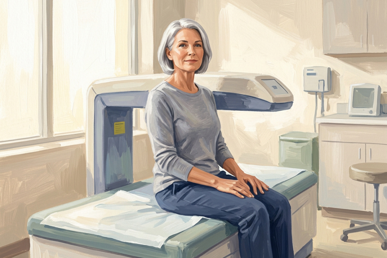
Diagnostic Standards: DXA Scans, T-Scores, and the FRAX Tool
Because bone loss is asymptomatic, healthcare providers rely on Dual-Energy X-ray Absorptiometry (DXA) scans to measure bone mineral density (BMD). A DXA scan is a low-radiation imaging test, usually targeting the hip and lumbar spine, which are the most reliable indicators of fracture risk.
The results are primarily reported as a T-score, which compares a woman's bone density to the peak bone mass of a healthy 30-year-old.
| T-Score Range | Clinical Classification |
|---|---|
| -1.0 or higher | Normal Bone Density |
| -1.0 to -2.5 | Osteopenia (Low Bone Mass) |
| -2.5 or lower | Osteoporosis |
| -2.5 or lower + Fracture | Severe (Established) Osteoporosis |
Providers may also look at Z-scores, which compare bone density to what is expected for someone of the same age, sex, and ethnicity. A Z-score of -2.0 or lower may suggest that something other than menopause — such as a medical condition or medication — is contributing to bone loss.
For more on what these results mean and how often DXA scans are covered, visit Groove Health to learn about Medicare-covered bone health programs.
Using FRAX to Assess Fracture Risk
A T-score alone does not tell the whole story. To assess the actual likelihood of a break, clinicians use the FRAX (Fracture Risk Assessment Tool). This tool calculates the 10-year probability of a major osteoporotic fracture by combining BMD data with other risk factors, including:
- Age and ethnicity
- History of previous fractures
- Parental history of hip fracture
- Smoking and excessive alcohol use
- Long-term use of glucocorticoids (like prednisone)
If your FRAX results show that your chance of breaking a hip in the next 10 years is more than 3%, or your chance of having a major osteoporosis-related fracture is 20% or higher, your clinician may recommend medication — even if your T-score has not yet reached -2.5. Guidance from the Endocrine Society notes that treating bone loss earlier, during osteopenia, can help lower the risk of progressing to full osteoporosis.
Nutritional Strategies for Bone Preservation
Nutrition provides the raw materials for bone maintenance. For postmenopausal women, the goal is to ensure the body has sufficient minerals to support the remodeling process. For a deeper look at building a bone-friendly diet, see our guide on nutrition for bone health.
- Calcium: The Bone Health & Osteoporosis Foundation (BHOF) recommends 1,200 mg of calcium daily for women over 50. It is best to obtain this through diet, as food-based calcium is absorbed more efficiently. Sources include dairy products, fortified plant milks, sardines (with bones), and dark leafy greens like kale or bok choy. Our supplement guide for bone density covers when and how to supplement safely.
- Vitamin D: Without Vitamin D, the body cannot absorb calcium. Most experts recommend 800–1,000 IU of Vitamin D3 daily. Since it is difficult to get enough through food or sunlight alone, supplements are often necessary, especially for those in northern climates or who spend most of their time indoors.
- Protein: Bone is roughly 50% protein by volume. Adequate protein intake is vital for maintaining both bone density and the muscle mass needed to support the skeleton and prevent falls. Learn more in Building Bones: Why Protein Matters.
Lifestyle adjustments matter too. Smoking is directly toxic to bone-building cells and accelerates estrogen clearance from the body. Limiting alcohol to no more than one drink per day helps prevent interference with calcium absorption and reduces the risk of falls.
Exercise That Strengthens Bones
Exercise is "mechanical medicine" for the skeleton. Bones respond to physical stress by becoming denser and stronger — a principle known as Wolff's Law. For a complete breakdown of which exercises are most effective, see The Best Exercise for Bones.
- Weight-Bearing Aerobic Exercise: Activities where the body works against gravity — walking, jogging, dancing, and pickleball. Research indicates these should be performed for at least 30 minutes, most days of the week. Our article on how exercise prevents osteoporosis progression covers the evidence in detail.
- Resistance Training: Using weights, bands, or body weight (like squats or wall push-ups) to pull on the bone via muscle contraction, stimulating osteoblast activity. Strength Training and Osteoporosis is a good starting point if you are new to lifting.
- Balance and Posture: Tai Chi or specific physical therapy drills improve proprioception. This is vital because the best way to treat a fracture is to prevent the fall that causes it.
For those unsure where to start, you can check your eligibility for personalized exercise plans through Groove Health, which pairs patients with physical therapists to build safe, effective home routines.
Pharmacological Management: Hormone Therapy and Bone Medications
When lifestyle changes are not enough, several classes of medication can help manage postmenopausal bone loss.
- Menopausal Hormone Therapy (MHT): In women within 10 years of menopause or under age 60, MHT is a highly effective first-line option. By replacing lost estrogen, it directly addresses the root cause of postmenopausal bone loss.
- Bisphosphonates: The most commonly prescribed osteoporosis drugs (e.g., Alendronate, Zoledronic acid). These are "antiresorptive" agents that slow osteoclast activity, allowing bone-building cells to catch up.
- Denosumab: A RANKL inhibitor administered as an injection every six months. It prevents the maturation of osteoclasts and is highly effective, but requires strict adherence — bone loss can rebound rapidly if a dose is missed.
- SERMs (Selective Estrogen Receptor Modulators): Drugs like Raloxifene act like estrogen in bones but block its effects in breast tissue, offering a dual benefit for some women.
Anabolic vs. Antiresorptive Treatments
For women at very high risk — such as those who have already had a fracture or have very low T-scores — clinicians may recommend anabolic (bone-building) medications.
Unlike bisphosphonates, which slow breakdown, anabolics like Teriparatide or Romosozumab stimulate new bone formation. Clinical evidence suggests that for high-risk patients, starting with an anabolic agent for one to two years and then "locking in" those gains with an antiresorptive provides the strongest protection against fractures.
Long-Term Fracture Prevention and Fall Risk Mitigation
The ultimate goal of managing osteoporosis is preserving independence. A hip fracture can be life-altering: statistics show that 20–30% of patients over 65 die within a year following a hip fracture due to complications like pneumonia or blood clots.
Beyond medication and exercise, fall-proofing the home environment is a critical step:
- Lighting: Ensure hallways and bathrooms are well-lit at night.
- Obstacles: Remove throw rugs and clear clutter from walking paths.
- Vision and Hearing: Regular check-ups help maintain the sensory input needed for balance.
- Gait Assessment: A physical therapist can identify subtle changes in walking patterns that may increase trip risk.
For a practical checklist covering both movement and diet, see A Starter Guide for Movement & Food.
Frequently Asked Questions About Menopause and Bone Loss
When should I get my first bone density scan?
The general clinical guideline is age 65 for all women. However, women who enter menopause early (before age 45), have a history of smoking, weigh less than 127 pounds, or have a family history of hip fractures should request a baseline DXA scan at the onset of menopause.
Can I reverse bone loss after menopause?
While it is difficult to completely reverse osteoporosis back to normal bone density, modern treatments — especially anabolic medications combined with antiresorptives — can significantly increase bone mineral density and, more importantly, strengthen the bone's internal architecture to reduce fracture risk.
Is hormone replacement therapy safe for my bones?
For most healthy women under 60 who are within 10 years of menopause, the benefits of MHT for bone protection often outweigh the risks. This is known as the "Window of Opportunity" hypothesis. Safety depends on individual health history, so a consultation with your healthcare provider is essential.
Taking the Next Step
Understanding how menopause causes osteoporosis is the first step toward a proactive strategy that prioritizes strength, stability, and long-term independence. Groove Health provides a comprehensive, Medicare-covered approach to managing postmenopausal bone loss — pairing patients with a physician and a dedicated physical therapist for personalized care plans.
If you are ready to get started, Groove's Osteoporosis Starter Guide walks you through what to expect, or you can check your eligibility directly.
Works Cited
- Bone Health & Osteoporosis Foundation. "What Women Need to Know." Bone Health & Osteoporosis Foundation (BHOF) Patient Education Resource, 2023.
- Neer RM. "Bone Loss Across the Menopausal Transition." Annals of the New York Academy of Sciences, 2010.
- Xiong J, Onal M, Jilka RL, Weinstein RS, Manolagas SC, O'Brien CA. "Estrogen Regulates Bone Turnover by Targeting RANKL Expression in Bone Lining Cells." Scientific Reports, 2017.
- Greendale GA, Sowers M, Han W, Huang MH, Finkelstein JS, Crandall CJ, Lee JS, Karlamangla AS. "Bone Mineral Density Loss in Relation to the Final Menstrual Period in a Multiethnic Cohort: Results from the Study of Women's Health Across the Nation (SWAN)." Journal of Bone and Mineral Research, 2012.
- World Health Organization Scientific Group. "Assessment of Osteoporosis at the Primary Health Care Level: Report of a WHO Scientific Group." WHO Technical Report Series (WHO Press, Geneva), 2007.
- Kanis JA, Johnell O, Oden A, Johansson H, McCloskey E. "FRAX and the Assessment of Fracture Probability in Men and Women from the UK." Osteoporosis International, 2008.
- Cosman F, de Beur SJ, LeBoff MS, Lewiecki EM, Tanner B, Randall S, Lindsay R; National Osteoporosis Foundation. "Clinician's Guide to Prevention and Treatment of Osteoporosis." Osteoporosis International, 2014.
- Haentjens P, Magaziner J, Colon-Emeric CS, Vanderschueren D, Milisen K, Velkeniers B, Boonen S. "Meta-analysis: Excess Mortality After Hip Fracture Among Older Women and Men." Annals of Internal Medicine, 2010.
- Ruff C, Holt B, Trinkaus E. "A 2003 Update of Bone Physiology and Wolff's Law for Clinicians." Journal of Orthopaedic and Sports Physical Therapy, 2004.
- Rossouw JE, Prentice RL, Manson JE, Wu L, Barad D, Barnabei VM, Ko M, LaCroix AZ, Margolis KL, Stefanick ML. "A 'Window of Opportunity': The Reduction of Coronary Heart Disease and Total Mortality with Menopausal Therapies is Age and Time Dependent." Menopause, 2007.
- Eghbali-Fatourechi G, Khosla S, Sanyal A, Boyle WJ, Lacey DL, Riggs BL. "Role of RANK Ligand in Mediating Increased Bone Resorption in Early Postmenopausal Women." Journal of Clinical Investigation, 2003.
- Bliuc D, Nguyen ND, Milch VE, Nguyen TV, Eisman JA, Center JR. "Mortality Risk Associated with Low-Trauma Osteoporotic Fracture and Subsequent Fracture in Men and Women." JAMA, 2009.
This content is for informational purposes only and does not constitute medical advice. Consult a qualified healthcare professional for diagnosis and treatment. If you are on Medicare and interested in a personalized bone health program, you can check your eligibility at groovehealth.com.